
Figure 8. C- and B-helix features in the bound and apo states.
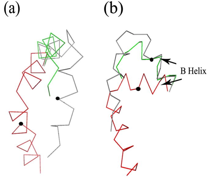
a) Three different αC helix arrangements: cAMP-bound wild type (red), partially unwound (R348A in gray) and disordered (R307W in green). The circle indicates the position of Phe 341, a residue referred in the text (the R307W structure lacks this residue). b) Three examples of the αB helix arrangement emphasizing how it occupies two orientations: bound (wild type in red) and apo (R348A in gray and R307W in green). The circle indicates the position of Leu 330 (see text).