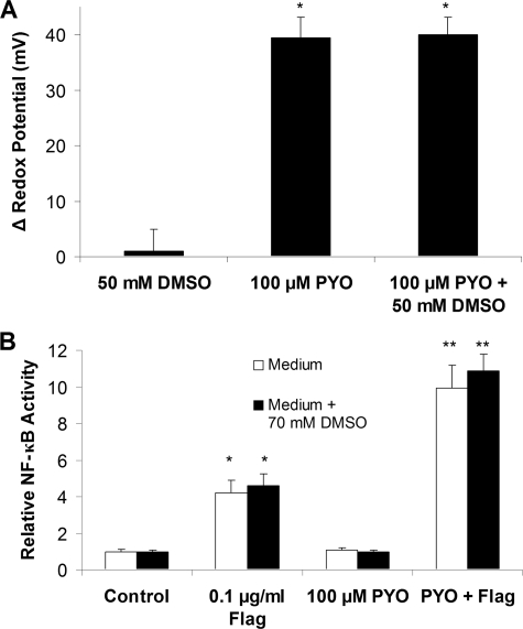
FIGURE 6.
Effects of DMSO on Ψcyto and NF-κB activity in cells treated with PYO and/or flagellin. A, CF15 cells expressing roGFP1 were treated with 50 mm DMSO, 100 μm PYO, and 100 μm PYO + 50 mm DMSO. Ψcyto were recorded before and after 2 h in five different regions of the cover glass each containing ≥10 different roGFP1-expressing cells. Averages ± S.D. for changes in Ψcyto in response to PYO/DMSO are shown; *, p < 0.05 compared with nontreated control. B, summary of effects of 100 μm PYO ± 70 mm DMSO in the absence or presence of flagellin (Flag) (10-7 g/ml) on NF-κB activity (expressed relative to controls = 1.0). Averages ± S.D. *, p < 0.05 compared with control; **, p < 0.05 compared with flagellin.

