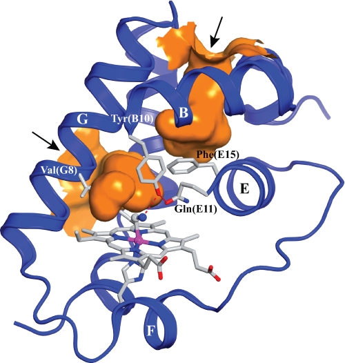
FIGURE 1.
View of the distal heme pocket and the tunnels of cyanomet-trHbN chain B under xenon pressure (PDB entry 1S56). Besides the protein backbone (blue ribbon, with labeled α-helices), the figure shows hydrogen-bonding (dashed red lines) between the distal residues Tyr(B10) and Gln(E11) and the heme-bound cyanide. The path of the two tunnels is shown in orange. The short tunnel (∼8 Å) connects the heme distal site to the outer solvent space at a location comprised between the central region of the G and H helices (left in the figure). The long tunnel (∼20 Å) extends from the heme distal cavity to a solvent access site located between the inter-helical loops AB and GH (upper part of the figure); note the gating role of Phe(E15) on the long tunnel. The arrows point to the tunnel entrance sites facing the solvent. The figure was produced using the PyMOL software (Delano Scientific).

