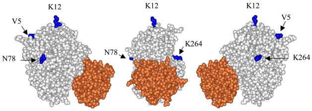
Figure 1.
The yeast iso-1 cytochrome c/CcP complex (9). Yeast cytochrome c is shown in red, CcP is shown in white and the residues in CcP that were individually converted to cysteine residues are shown in blue. Center - space-filling rendering of the complex with the cytochrome c in front of the CcP molecule. This view defines the ‘front face’ of CcP. The primary cytochrome c-binding site is located on the lower half of the front face of CcP. Three of the four CcP residues that are converted to cysteine residues can be seen in this view: Lys-12, Asn-78, and Lys-264. Right-hand side - A 90° clockwise rotation of the central figure about the vertical axis exposes the right face of the CcP molecule. Val-5, on the back side of CcP can be seen as well as the relative locations of Lys-12 and Lys-264. Left-hand side - A 90° counter-clockwise rotation of the central figure about the vertical axis exposes the left face of CcP. Val-5, Lys-12, and Asn-78 are seen in this view.

