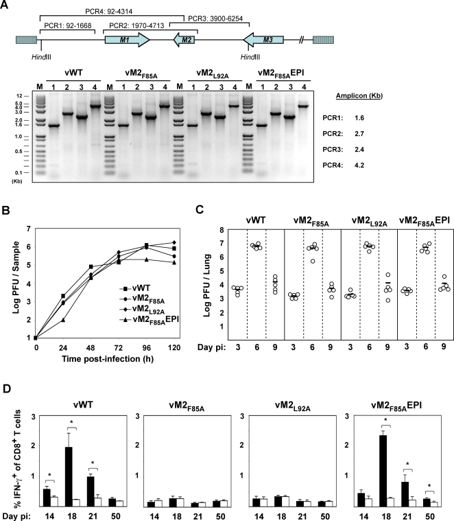
Figure 2. MuHV-4 latent epitope mutants show normal in vitro and in vivo replication.
A. High molecular weight DNA from MuHV-4-infected BHK-21 cells was checked by PCR for genome integrity in the HinDIII-E region. A schematic representation of the MuHV-4 genome and amplicon coordinates for each PCR is shown. B. BHK-21 cells were infected (0.01 PFU per cell) with the indicated viruses, washed in PBS and virus replication with time was monitored by plaque assay. C. BALB/c mice were intranasally infected with 104 PFU of the indicated viruses. At the indicated days post-infection lungs were removed and titrated for infectious virus by plaque assay. Each point represents the titre of an individual mouse. Horizontal lines indicate arithmetic means. None of the mutants showed a deficit relative to the wild-type (p>0.5 by 1-way non-parametric ANOVA Kruskal-Wallis Test). D. Splenocytes of BALB/c mice infected with viruses of the indicated genotypes were stimulated in vitro with either M284–92 (black bars) or EGFP200–208 as a control (white bars) in the presence of Brefeldin A, then stained for intracellular interferon-gamma. The data show the percentage of CD8+ T cells responding to peptide at each time point (arithmetic means±SEMs from 3 independent measurements). *, p<0.05 using a 1 tailed Student's t-test.

