
Figure 1. Protein and dsRNA Gels of PsV-S and PsV-F.
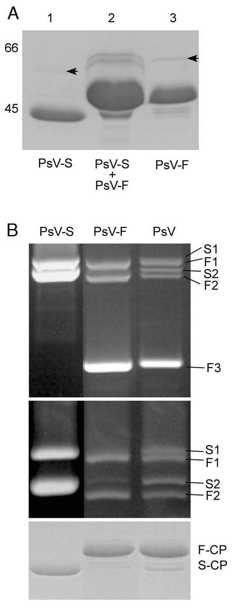
Top panel: SDS/PAGE analysis of ion-exchange chromatography fractions. Lane 1, peak 1 contained only PsV-S; lane 2, peak 2 contained a mixture of PsV-S and -F; lane 3, peak 3 was highly enriched in PsV-F and contained only traces of PsV-S. The relative positions of 66- and 45-kDa markers (data not shown) are indicated to the left of lane 1. The virus preparation analyzed in this panel was independent from that analyzed in the middle and bottom panels.
Middle panels: Agarose-gel electrophoresis of virion dsRNA isolated from ion-exchange-purified PsV-S and PsV-F as well as the unfractionated gradient-purified mixture of both viruses. The agarose gel was run for 1 h (upper) or 3 h (lower). S1 and S2 correspond to PsV-S dsRNA1 and dsRNA2, respectively; F1, F2, and F3 correspond to PsV-F dsRNA1, dsRNA2 and satellite dsRNA, respectively.
Bottom panel: SDS/PAGE analysis of the CPs of PsV-S and -F, purified by ion-exchange chromatography, as well as of the unfractionated gradient-purified PsV preparation. F-CP, PsV-F capsid protein; S-CP, PsV-S capsid protein.