
Figure 5.
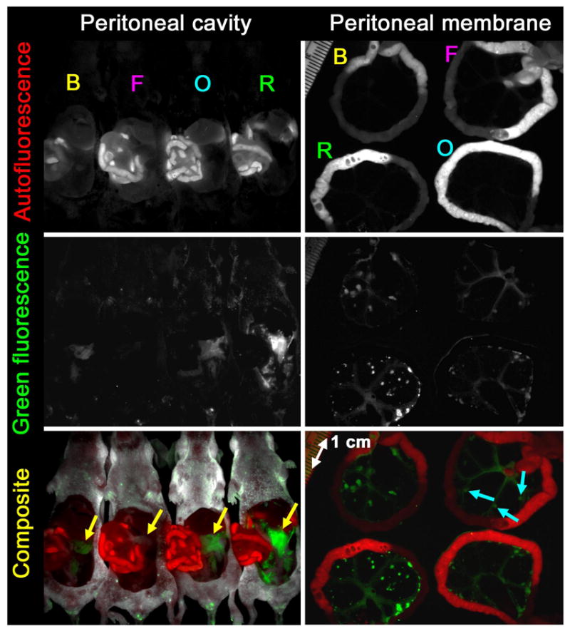
In vivo spectral fluorescence images of tumor-bearing mice 3h after intraperitoneal injection with Av-BODIPY (B), Av-FITC (F), Av-OreG (O) and Av-RhodG (R). Upper: Autofluorescence image. Middle: Green dye fluorescence image. Lower: Composite image (red: autofluorescence, green: green dye fluorescence). Spectral fluorescence image of the peritoneal cavities clearly visualized the disseminated tumor foci (yellow arrows) in mice incubated with Av-BODIPY, Av-OreG or Av-RhodG whereas Av-FITC failed to visualize tumor foci (yellow arrow) due to insufficient fluorescence intensity. Closeup image of the peritoneal membranes demonstrates peritoneal implants histologically confirmed to be metastatic deposits as small as 1 mm in diameter in all mice including Av-FITC injected mouse (blue arrows). The fluorescence intensity of Av-RhodG was the highest and that of Av-FITC was the lowest of all when compared visually.