
Fig. 1.
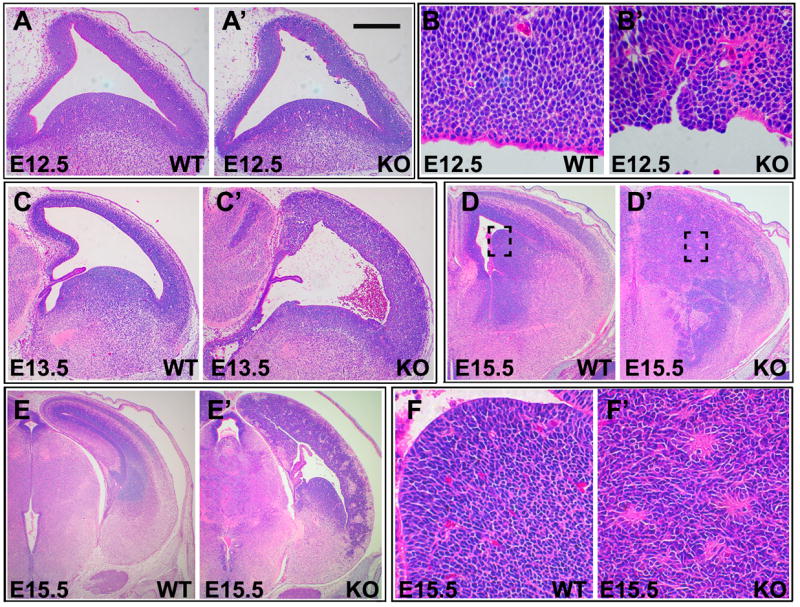
Severe dysplasia and hyperplasia in αE-catenin−/− brain. Histologic appearance of brains from wildtype (WT) and αE-cateninLoxP/LoxP/Nestin-Cre+/− (KO) mice. Sagittal sections through developing telencephalon from the wildtype (A, C) and αE-catenin−/− (A′, C′) brains of E12.5 (A, A′) and E13.5 (C, C′) embryos. Ventricular zone of the cerebral cortex from the E12.5 wildtype (B) and αE-catenin−/− (B′) brains. Coronal sections from the E15.5 wildtype (D, E, F) and αE-catenin−/− (D′, E′, F′) brains. Areas in dashed squares in D, D′ are shown at higher magnification in F, F′. Bar in A′ represents 0.27 mm in A, A′; 0.36 mm in C–C′; 0.42 mm in D, D′; 0.54 mm in E–E′; 50 km in F, F′, 40 km in B, B′.