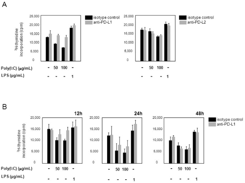
Figure 5. Blockade of PD-L1 restores CD4+ T-cell proliferation.

(A) DCs were stimulated with poly(I:C) or LPS for 5 hours and incubated with anti-PD-L1- (1 µg/mL) (left panel), or anti-PD-L2- (10 µg/mL) (right panel) blocking mAb, or the appropriate isotype control for 1 hour. 1×10⁵ CD4+ T cells were added, and proliferation was assessed by [3H]thymidine incorporation 72 hours later. One representative experiment of three is shown. Left panel: Unstimulated DC versus poly(I:C)-stimulated (100 µg/mL) DC, P = 0.0003; unstimulated DC versus LPS-stimulated (1 µg/mL) DC, P = 0.008; poly(I:C)-stimulated (100 µg/mL) DC versus mAb-blocked poly(I:C)-stimulated (100 µg/mL) DC, P = 0.01. Right panel: Unstimulated DC versus poly(I:C)-stimulated (100 µg/mL) DC, P = 0.03; unstimulated DC versus LPS-stimulated (1 µg/mL) DC, P = 0.009. (B) Anti-PD-L1 blocking mAb was added at different time points after initiation of T cell-DC interaction, and T-cell proliferative responses were measured as described above. One representative experiment of three is shown. 12 hours: Unstimulated DC versus poly(I:C)-stimulated (100 µg/mL) DC, P = 0.04; poly(I:C)-stimulated (100 µg/mL) DC versus mAb-blocked poly(I:C)-stimulated (100 µg/mL) DC, P = 0.03. 24 hours: Unstimulated DC versus poly(I:C)-stimulated (100 µg/mL) DC, P = 0.01. 48 hours: Unstimulated DC versus poly(I:C)-stimulated (100 µg/mL) DC, P = 0.05.
