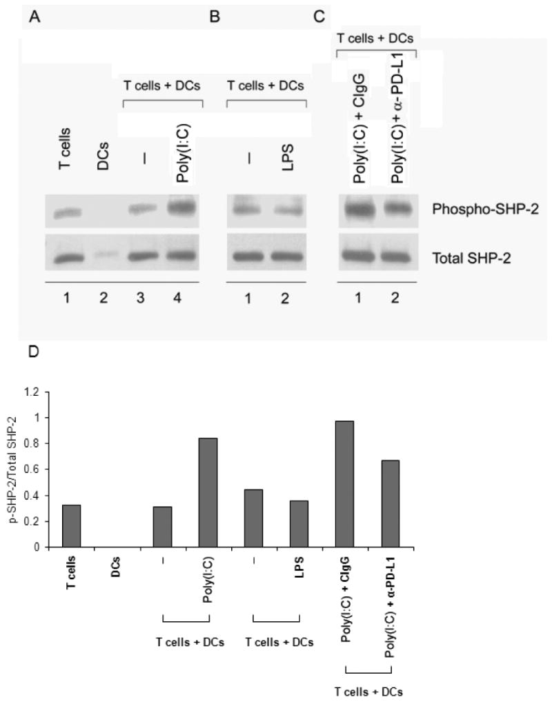
Figure 6. TLR3-stimulated DCs enhance SHP-2 phosphorylation in CD4+ T cells downstream of PD-1/PD-L1 interaction.

(A, B) CD4+ T cells were cocultured with unstimulated (A, lane 3 and B, lane 1), poly(I:C)-pretreated (A, lane 4), or LPS-pretreated (B, lane 2) DCs for 24 hours, lysed, and cell extracts were resolved on SDS-PAGE acrylamide gels. Proteins were transferred onto PVDF membranes and membranes probed for phospho-SHP-2 (upper row). Membranes were stripped and reprobed for total SHP-2 (lower row). Lanes 1 and 2 in (A) are T cells and DCs cultured alone. (C) CD4+ T cells were cocultured with DCs as in (A), lane 4, but with the addition of either control-IgG1 (lane 1) or anti-PD-L1 blocking mAb (lane 2) and probed for phospho- and total SHP-2. Results in (A), (B), and (C) are representative of three independent experiments. (D) Quantification of the phospho-SHP-2/SHP-2 ratios was performed using the ImageJ software (NIH).
