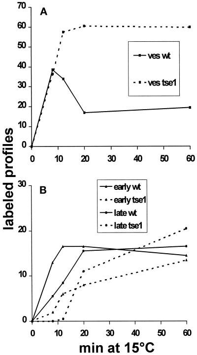
Figure 8.
Quantitation of Nanogold-labeled structures. Wild-type and tlg2Δ spheroplasts were incubated with positively charged Nanogold on ice for 5 min and then shifted to 15°C for various times up to 60 min. The cells were fixed, dehydrated, and embedded. Thin sections were generated and enhanced with HQ Silver and visualized in the electron microscope. Labeled vesicles, early endosomes, and late endosomes were identified on sections and quantified. The vesicle quantitation is shown in A, the early and late endosome quantitation in B.

