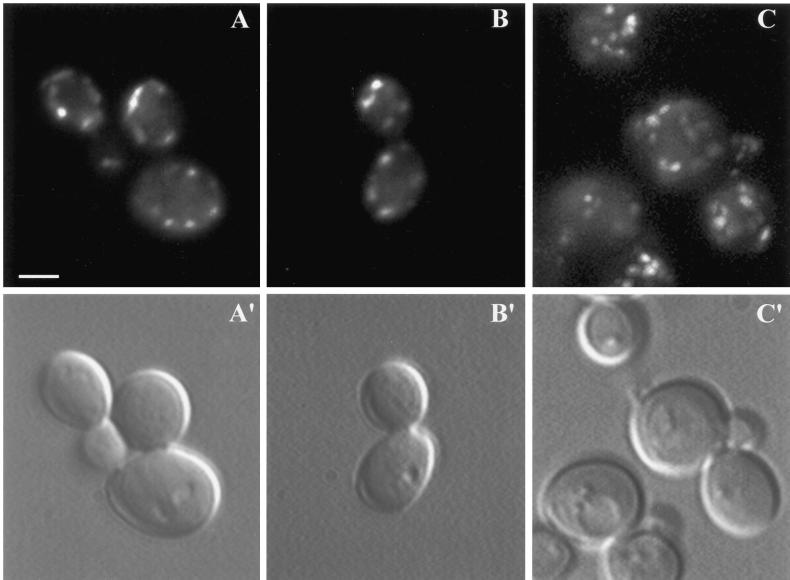
Figure 9.
Localization of GFP-tagged Tlg2p and Sec7p. Fluorescence of GFP-tagged Tlg2p in the presence of 1 mM methionine is shown to the left (A and B), and fluorescence of GFP-tagged Sec7p is shown to the right (C); the corresponding cells visualized with DIC optics are shown below (A′–C′). In cells expressing GFP-tagged Tlg2p, the fluorescence is located in small structures at the periphery of the cells, not linked with the vacuole. Twenty-seven of 35 cells observed presented the same fluorescence pattern. In cells expressing GFP-tagged Sec7p, the fluorescence is located in punctate structures throughout the cytoplasm. All the cells observed (33) presented the same pattern. Bar, 2.5 μm.

