
Figure 3.
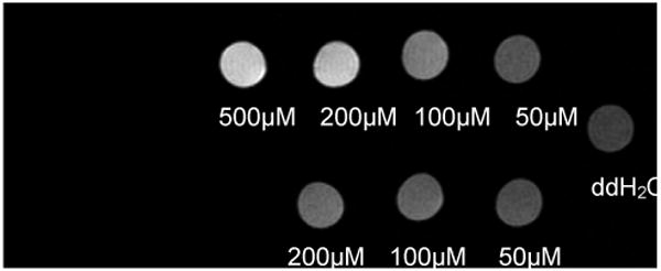
Mal-BSA (Gd-DOTA)n is taken up by macrophages. P388D1 cells were incubated with n = 15 (top row) or n = 10 (bottom row) mal-BSA (Gd-DOTA)n for one hour then imaged at 7T. Contrast increases in a concentration dependent manner (right to left) indicating uptake of the contrast agent by the cells. Contrast enhancement is observed for an application of as low as 50μM contrast agent to the cells (top row, rightmost column). Actual T1 values were, left-right: (top row) = 0.84s, 0.96s, 1.49s. 2.33s; (bottom row) = 1.79s, 1.96s, 2.51s; (water) = 3.44s