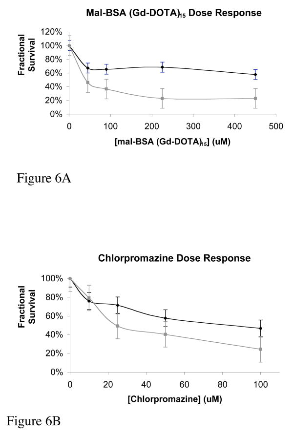
Figure 6.
Toxicity of mal-BSA. Cytotoxicity was assessed for mal-BSA (Gd-DOTA)15 and chlorpromazine as a positive control. The assays were carried out in parallel and in triplicate. Cells were counted and plated evenly in a 96-well plate. After a 24-hour adhesion time, varying concentrations of mal-BSA (Gd-DOTA)15 (0, 45, 90, 225, and 445μM) or chlorpromazine (0, 10, 25, 50, and 100μM) were placed on the cells. After a 4- and 24-hour agent exposure time at 37°C the MTT assay was performed. Dose response curves at 4- (black) and 24- (gray) hours are shown for mal-BSA (Gd-DOTA)15 (Figure 6A) and chlorpromazine (Figure 6B). For 4h mal-BSA incubation, toxic dose EC50 is not reached for the concentrations of agent tested (EC50 > 450μM). For the 24h mal-BSA EC50 = 40μM. This is in contrast with chlorpromazine with EC50 = 84μM for 4h incubation and EC50 = 24μM for 24h incubation. Toxicity of mal-BSA (Gd-DOTA) is less that that of the chlorpromazine standard. Error bars reflect standard error.

