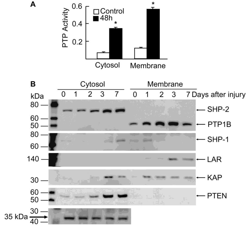
Figure 1. PTP activity and expression during “in vivo” epithelial wound healing.

New Zealand rabbits were injured by gently scraping the corneas, leaving the limbal epithelium intact. Cornea epithelium from control and injured rabbits was collected and cytosol and membrane fractions prepared as explained in Methods. A) Total PTP activity was measured with 5 µg protein as explained in Methods before and after 48 h injury. Values represent the average ± SD of three samples. * significant differences with respect to control. B) Samples were collected 1, 2, 3 and 7 days after the injury and the same amount of proteins were separated by SDS polyacrilamide gel electrophoresis and analyzed by immunoblotting with PTPs antibodies. As loading controls for the cytosol fraction, a 35 kDa protein that appears in the gels after blotting with some of the antibodies, and did not change its expression during would healing, was used. In the membrane fraction, the phosphatase KAP also exhibited no change between 1–7 days after injury. The data represent one of two experiments.
