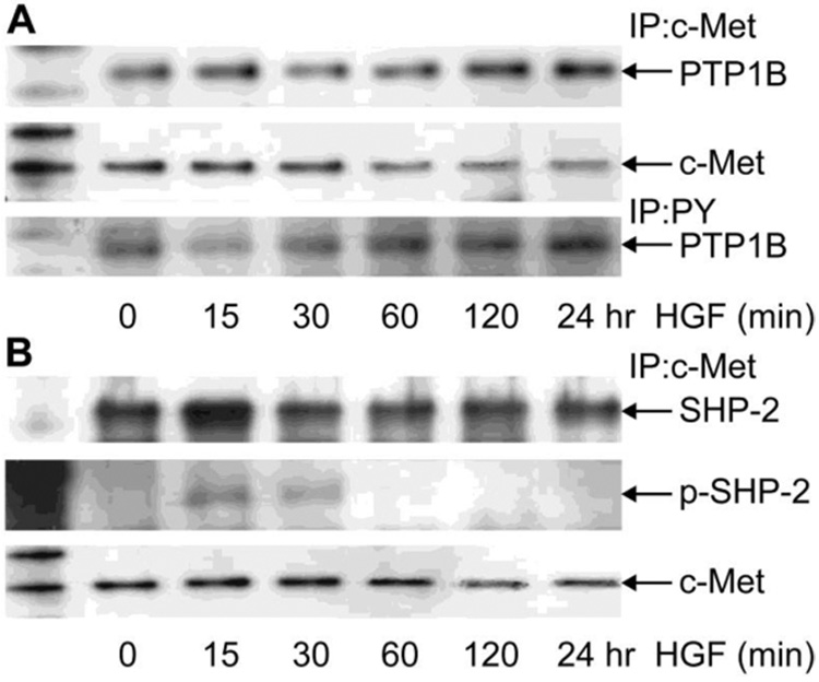
Figure 4. Bound to c-Met and phosphotyrosine content of PTP1B and SHP2.

HCE cells were incubated in the absence or presence of 40 ng/ml HGF for different times and then collected in lysis buffer. Cell lysate (1mg) was immunoprecipitated with monoclonal c-Met antibody (A and B) or with PY monoclonal antibody (A) as indicated in Methods. Proteins were separated by 4–12 % gradient gels and transferred to PVDF membranes. The membranes were immunoblotted with the polyclonal PTP1B antibody (A) or with the polyclonal SHP-2 and the phosphorylated polyclonal p-SHP-2 antibody (B). The membranes were stripped and reprobed with c-Met antibody. The data represent one of three similar experiments.
