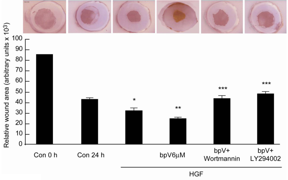
Figure 7. bpV(phen) increases HGF-stimulated epithelial wound healing that is reversed by PI-3K inhibitors.

Corneas were injured and cultured in a serum free medium for 24 h in the presence of HGF (40 ng/ml) with or without 6 µM bpV( phen) or with or without 200 nM wortmannin or 20 µM LY294002. The corneas were stained with Alizarin red and the remaining uncovered area calculated. The pictures represent a cornea in each condition. The bars correspond to 5–6 corneas in each treatment. *p<0.05 compared to control at 24 h, **p< 0.05 compared to HGF, ***p< 0.05 compare to bpV(phen) + HGF (Student test analysis).
