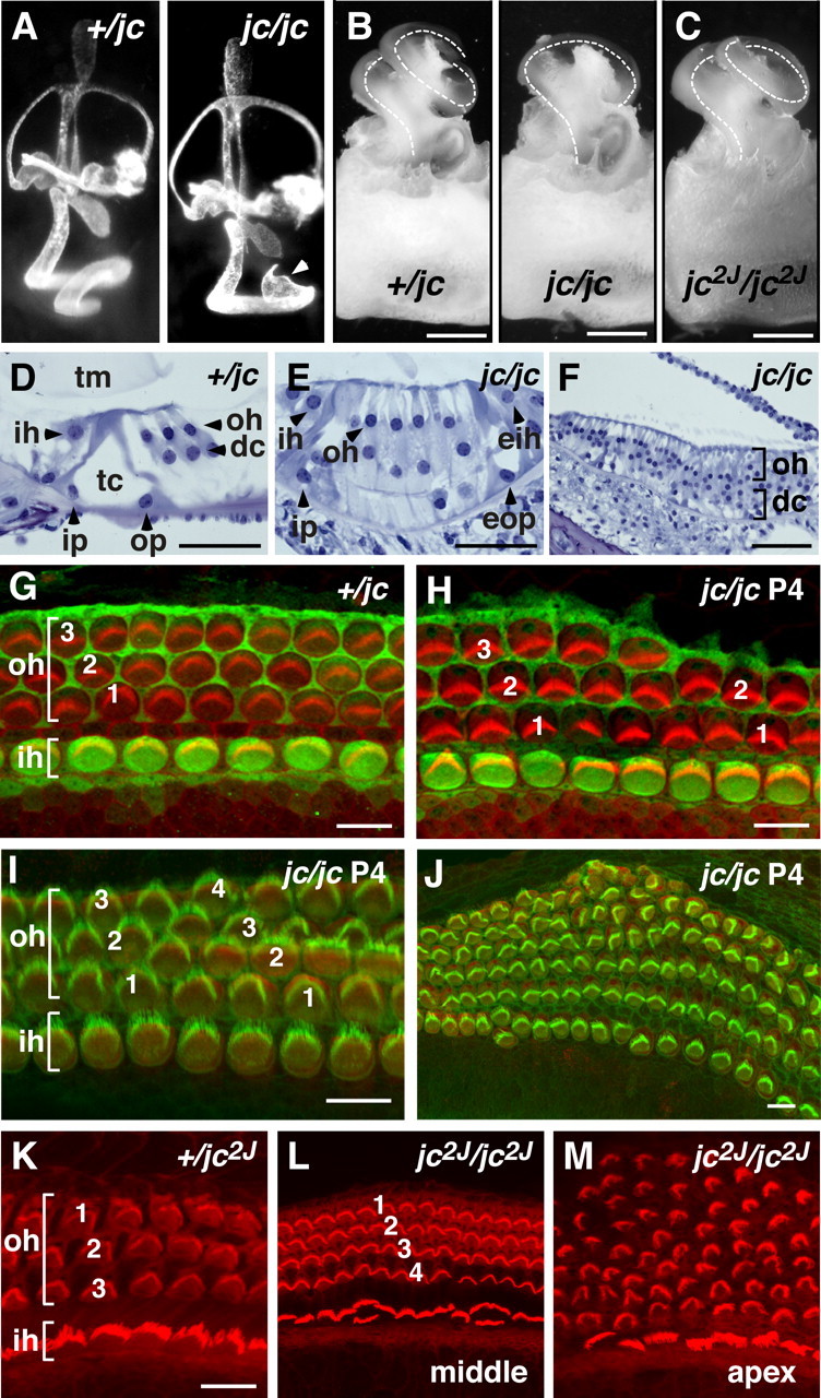
Figure 4.

Cochlea dysplasia in jc mutants. A, Inner ears injected with latex paint of +/jc and jc/jc at embryonic day 15.5. Note the bulbous structure at the apex of the shortened cochlea (arrowhead). B, C, Cochlear ducts from P12 (+/jc, jc/jc) and P14 (jc2J/jc2J) mice are shown. Note that the cochlear duct in the jc2J mutant is less shortened than in jc. The cochlear coil is indicated by the dotted lines. D–F, Plastic sections of the organ of Corti stained by toluidine blue O are shown. The organ of Corti is normally developed in 3-week-old +/jc mice (D). The locations of IHCs (ih), OHCs (oh), Deiter's cells (dc), inner (ip) and outer (op) pillar cells, tectorial membrane (tm), and tunnel of Corti (tc) are depicted. A section from the midapical region of a jc shows four rows of OHCs (E), an ectopic IHC (eih), and an ectopic outer pillar cell (eop) at the lateral edge. At the apical region, supernumerary rows of OHCs (oh, bracket) and Deiter's cells (dc, bracket) are seen (F). G–M, Confocal images of organ of Corti surface preparations. Staining with S100A1 (green) and phalloidin shows a normally developed organ of Corti in jc heterozygotes (G) with one row of IHCs (ih, bracket) and three rows of OHCs (oh, bracket). At midbase in jc homozygotes, short stretches of only two rows of OHCs appeared (H). Staining with an anti-myosin VI antibody (a marker of differentiated hair cells; red) identifies four rows of OHCs at the midapical region and up to six rows of OHCs at the more apical region (J). Phalloidin staining (red) of jc2J organ of Corti shows four rows of OHCs at the midapical region (compare +/jc2J with jc2J/jc2J; K, L) and a disorganized pattern of OHCs at the apex (M). Scale bars: A–C, 500 μm; D–F, 50 μm; G–K, 10 μm.
