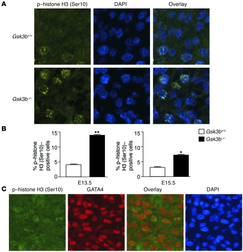
Figure 3. Increased cardiomyocyte proliferation in GSK-3β–deficient hearts.
(A) Immunofluorescence staining for nuclear phospho–histone H3 (Ser10) (left panels) in GSK-3β–deficient (bottom) compared with WT (top) animals. Merging of the histone and the DAPI (middle) stains confirmed nuclear localization of the phospho–histone H3 (Ser10) staining (right panels). Original magnification, ×40. (B) Composite mean ± SEM percentage of phospho–histone H3 (Ser10)–staining cells at E13.5 and E15.5 in the myocardium of WT (white bars) and GSK-3β–deficient (back bars) animals.*P < 0.05, **P < 0.001 versus WT. (C) Coimmunostaining of heart sections with phospho–histone H3 and GATA4, a cardiomyocyte marker, demonstrating colocalization in the overlay. Thus, the phospho–histone H3 (Ser10)–positive cells in the myocardium are overwhelmingly GATA4 positive, consistent with cardiomyocytes. Also shown is nuclear staining with DAPI. Original magnification, ×40.

