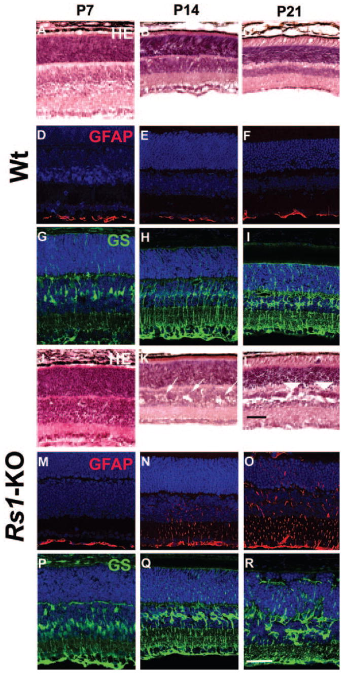
Figure 3.

Retinal morphology and glial cell immunohistochemistry of Wt and Rs1-KO during postnatal development. Hematoxylin and eosin–stained histology of Wt (A–C) and Rs1-KO (J–L) retinas at P7, P14, and P21. At P7 (J) no abnormal features were observed in the Rs1-KO retina. By P14, several schisis cavities (arrows) were observed in the INL of Rs1-KO retinas (K). There were no major histologic abnormalities in the OPL. In the P21 Rs1-KO retina, the cavities were much more extensive in the INL and were also found in the OPL (arrowheads) (L). The vacuoles in the INL of Wt retinas at P7 are processing artifacts (A). Müller cells and GFAP immunohistochemical staining: Müller cells, stained with GS antibody were identified at developmental stages P7, P14, and P21 (Wt, G–I; Rs1-KO, P–R). In Wt retina, only the nerve fiber and ganglion cell layer showed GFAP labeling (D–F). In Rs1-KO retina, GFAP expression at P7 was also restricted to the nerve fiber and ganglion cell layer, but between P14 (N) and P21 (O) GFAP labeling gradually extended across all layers, which is suggestive of Müller cell hypertrophy and reactive gliosis. Scale bar, 50 μm.
