
Figure 5.
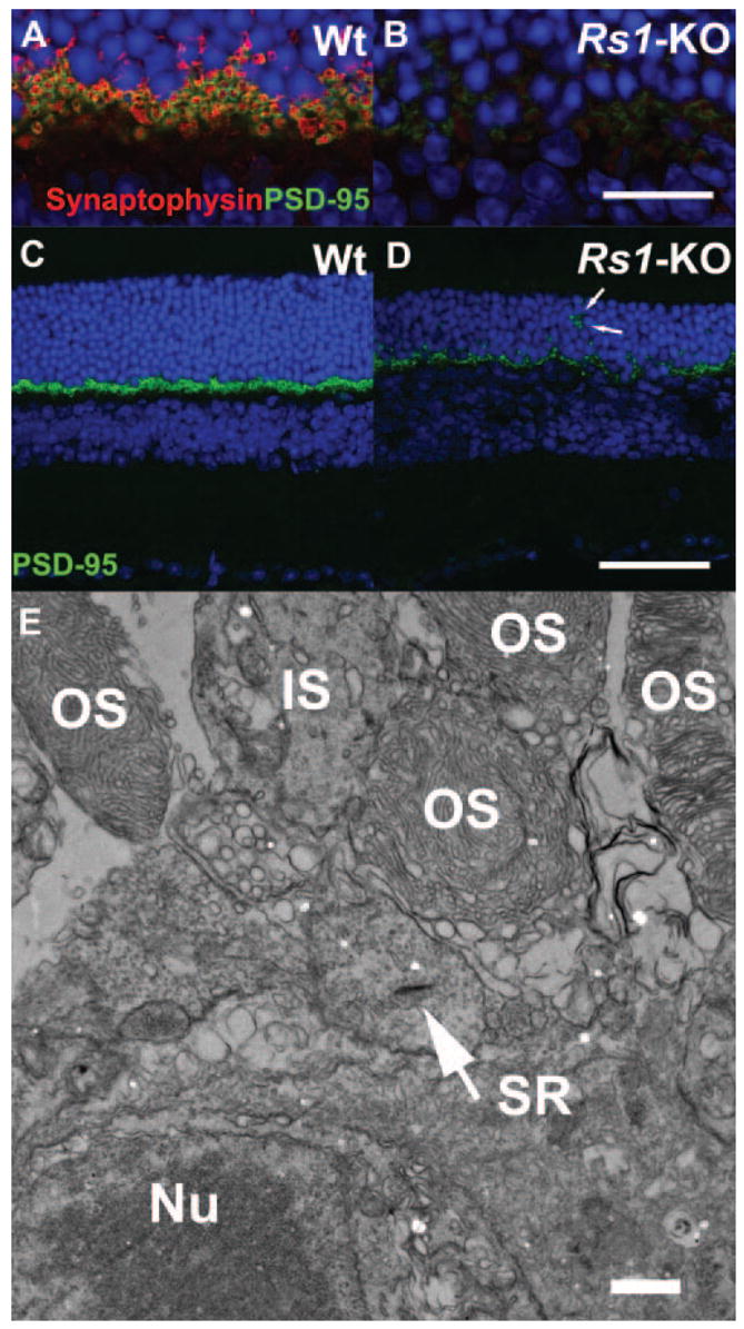
Double labeling of photoreceptor synaptic terminals in 4-month-old Wt and Rs1-KO retinas. Wt and Rs1-KO mouse retinas from 4-month-old mice were stained with the presynaptic marker proteins synaptophysin (marking synaptic vesicles, red) and PSD95 (green). Labeling of both markers was greatly decreased in Rs1-KO retina (B, D) compared with Wt (A, C). In Rs1-KO, some PSD95 label was located in the ONL (D, arrows) indicating displaced terminals. At the ultrastructural level (E), electron microscopy confirmed displacement of photoreceptor terminals containing synaptic ribbons (SR, arrow) between the outer segments and nuclei of photoreceptor cells. OS, outer segment; IS, inner segment; Nu, Nuclei. Scale bar: (A–D) 75 μm; (E) 100 nm.