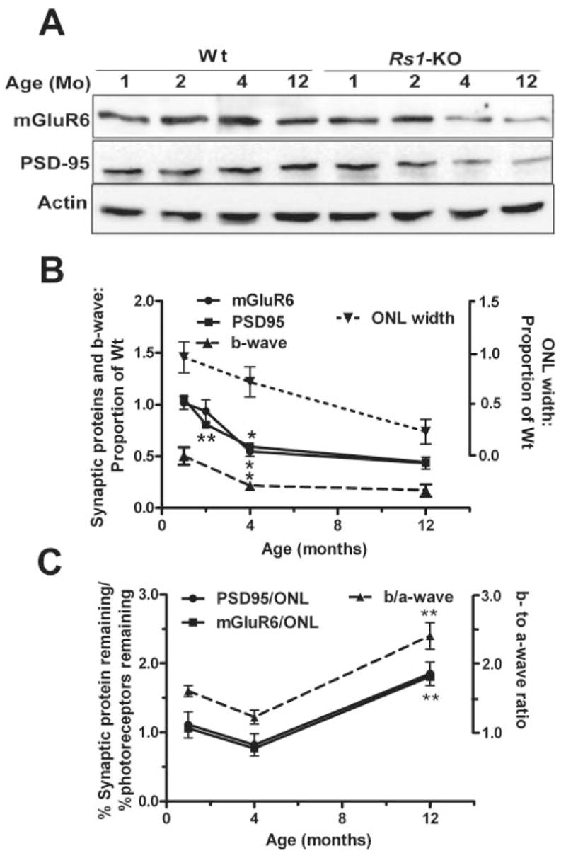
Figure 7.

Immunoblot analysis of aging changes in mGluR6 and PSD95 protein expression in Wt and Rs1-KO mice retinas. Representative Western blots from Wt and Rs1-KO mice of 1, 2, 4, and 12 months of age. The targeted proteins were detected and quantified using enhanced chemiluminescence. At 1 and 2 months of age Rs1-KO synaptic protein levels appeared comparable to Wt. Both PSD95 and mGluR6 protein levels declined across 2 to 12 months. (B) Protein levels as a proportion of Wt at each age (solid lines). Intensity of mGluR6 and PSD95 bands was normalized to β-actin band intensity, and the Rs1-KO value was divided by the Wt value at each age. The points were obtained from three independent experiments using a pool of two to three retinas from different mice in each experiment. The effect of age and genotype was analyzed by two-way ANOVA and Bonferroni posttest. Differences in synaptic protein levels between Wt and Rs1-KO retina were statistically significant at 4 and 12 months (P < 0.01). Significant change from the previous age (top asterisk at each point, mGluR6; bottom asterisk at each point, PSD95: *P < 0.05, **P < 0.01, one-way ANOVA, Bonferroni posttest). For comparison to previously reported retinal morphology and ERG studies, the ONL cell count and b-wave amplitude, normalized by Wt at each age, are plotted on the same graph (dashed lines). (C) Comparison of postsynaptic response (b-wave) to synaptic protein changes with age normalized by photoreceptor responses (a-wave) or number (ONL count), respectively. The ERG values are from a previous study.9 **Significant change from the previous age (P < 0.01).
