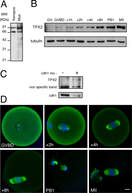
Figure 1. Characterization of TPX2 during mouse oocyte meiotic maturation.
A: Mouse oocytes (n = 200) and Xenopus metaphase II oocyte lysate (equivalent to 1/4 of oocyte) were immunoblotted using an anti-TPX2 antibody. The upper band corresponds to TPX2. The lower band is non-specific [11]. B: TPX2 accumulates progressively during meiotic maturation. For each time point, 140 mouse oocytes were immunoblotted using anti-TPX2 (only the TPX2 specific band is presented) and anti-α-tubulin antibodies. C: Cdh1 controls TPX2 levels at GV (Prophase I). Western blot analysis of TPX2 and cdh1 in control oocytes and cdh1 morpholino injected oocytes (n = 100 for each group). D: Localization of TPX2 during meiotic maturation. Oocytes were double stained for TPX2 (green) and nucleic acids (blue) at the times indicated relative to GVBD. In meiosis I and II, TPX2 decorates the spindle microtubules. Scale bar is 10 µm.

