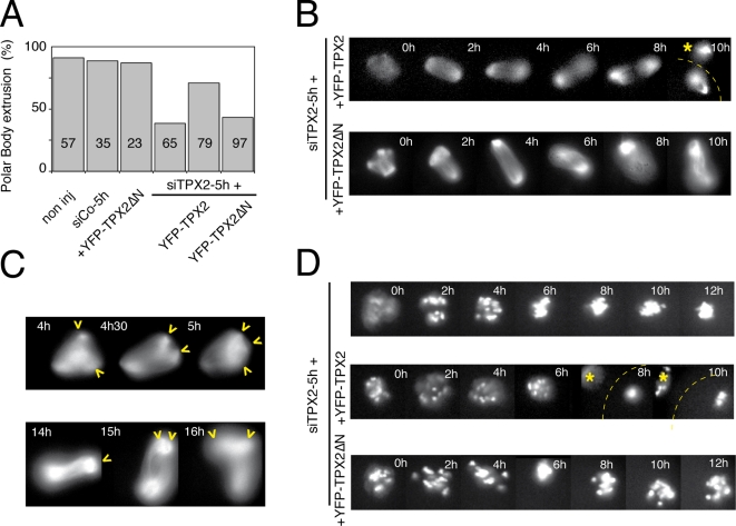
Figure 5. TPX2 activity relies on two distinct protein domains in the oocyte.
A: Percentage of polar body extrusion in different experimental conditions. Non-injected, siTPX2-5h, YFP-TPX2ΔN injected oocytes as well as oocytes co-injected with siTPX2-5h and YFP-TPX2 or YFPTPX2ΔN were analyzed. The total number of oocytes examined is indicated in each bar. B: Representative sequence of fluorescent images showing the microtubules organization in siTPX2-5h injected oocytes, which were co-injected either with YFP-TPX2 (upper panel) or YFP-TPX2ΔN (lower panel). Both YFP-TPX2 and YFP-TPX2ΔN rescued the spindle collapse. Asterisk indicates the polar body and the dotted line shows the outline of the oocyte. C: Representative fluorescent images of siTPX2-5h-injected oocytes co-injected with YFP-TPX2ΔN show spindle poles splitting (arrowheads). D: Representative sequence of fluorescent images showing the chromosomes siTPX2-5h injected oocytes expressing RFP-histone H2B alone (top panel), co-injected with either YFP-TPX2 (middle panel) or YFP-TPX2ΔN (bottom panel). The asterisk indicates the polar body and the dotted line shows the outline of the oocyte. Times are relative to the GVBD.

