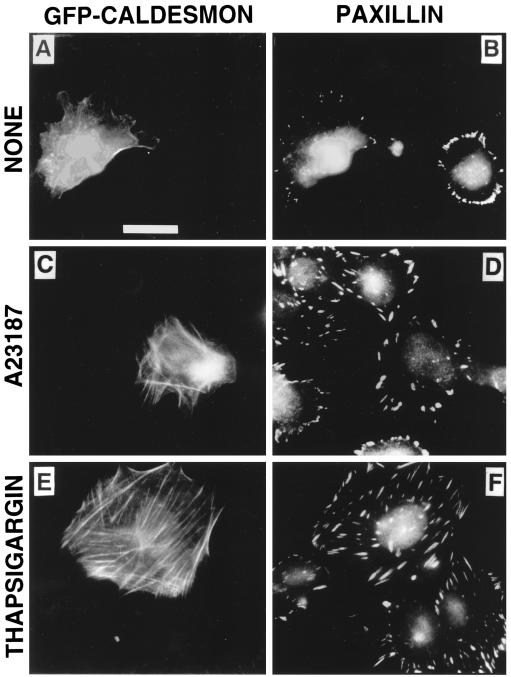
Figure 9.
Effect of caldesmon on the stimulation of focal adhesion formation by nocodazole. Cells were transfected with GFP–caldesmon and serum starved for 24 h before nocodazole treatment. After treatment the cells were fixed and stained with mouse monoclonal antibodies against paxillin. (A, C, and E) GFP–caldesmon fluorescence is visualized; (B, D, and F) staining of the same fields with anti-paxillin antibody is presented. Bar, 20 μm. (A and B) Incubation with 10 μM nocodazole for 30 min induced formation of prominent focal adhesions in nontransfected cells (B, right cell) but not in the cells transfected with caldesmon (B, left cell). (C–F) Increased intracellular Ca2+ interferes with the inhibitory effect of caldesmon on focal adhesion and stress fiber formation. Five micromolar A23187 (C and D) or 1 μM thapsigargin (E and F) was added to the medium for 1 h before incubation with 10 μM nocodazole for an additional 30 min. Note that incubation with either A23187 or thapsigargin restores the ability of caldesmon-transfected cells to form large focal adhesions and stress fibers after nocodazole treatment (compare B with D and F). Caldesmon in the cells treated with calcium-mobilizing agents is localized to stress fibers (C and E).

