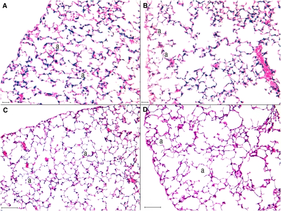
Figure 5.
Histologic samples of distal lung parenchyma. A represents alveolar parenchyma of 15-day-old nonexposed mice. In B, lung parenchyma of 15-day-old pre+postanally exposed mice. The alveolar spaces in B are enlarged when compared with those in A, as a result of incomplete alveolization. C represents distal alveolar parenchyma of 90-day-old nonexposed mice. The alveolar spaces are larger than in A and B. D shows the alveolar parenchyma of 90-day-old of pre+postanally exposed mice. The alveolar spaces are enlarged when compared with D. a = alveolar spaces. Hematoxylin and eosin staining. Scale bar: A, B = 20 μm; C, D = 100 μm.

