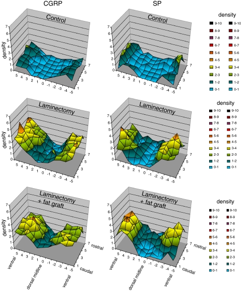
Fig. 2.
Three dimensional graphs showing the mean densities of CGRP- and SP-immunopositive nerve fibers per microscopical vision field in the dura of L4 of controls (group 1), laminectomized rats (group 2), and rats with laminectomy that received free autologous fat grafts (group 3). Six weeks post laminectomy the mean densities increased in ventral as well as in dorsal parts of the dura, independent of the use of fat grafts

