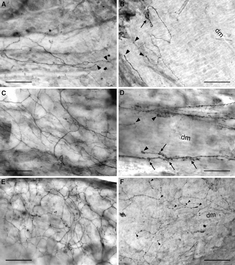
Fig. 3.
CGRP-immunopositive nerve fibers in the ventral (a, c, e) and dorsal (b, d, f) dura mater lumbalis of controls (a, b), laminectomized rats (c, d), and animals that underwent laminectomy and application of fat grafts (e, f); light microscopic micrographs of whole-mount preparations. a Thin bundles of nerve fibers and single neurons of small diameter innervating the ventral dura. Some of them terminate with straight and thickened end branches in the connective tissue (arrowheads). b Nerve fibers running in parallel to the dorsal midline and showing a coiled (arrow) and straight thickened terminals (arrowheads) in the connective tissue of the dorsal dura. c Dense network of immunopositive nerve fibers in the ventral dura mater lumbalis. d Small bundles of stained varicose nerve fibers branching and terminating (arrowheads) in the dorsal dura. Note the axonal spine-like structures (arrows) at the end branches. e Prominent network of immunopositive nerve fibers in the ventral dura. f Network of stained varicose nerve fibers arborizing and terminating in the connective tissue along the dorsal midline. Note the thickened nerve terminals (arrowheads). dm dorsal midline. Bars 100 μm

