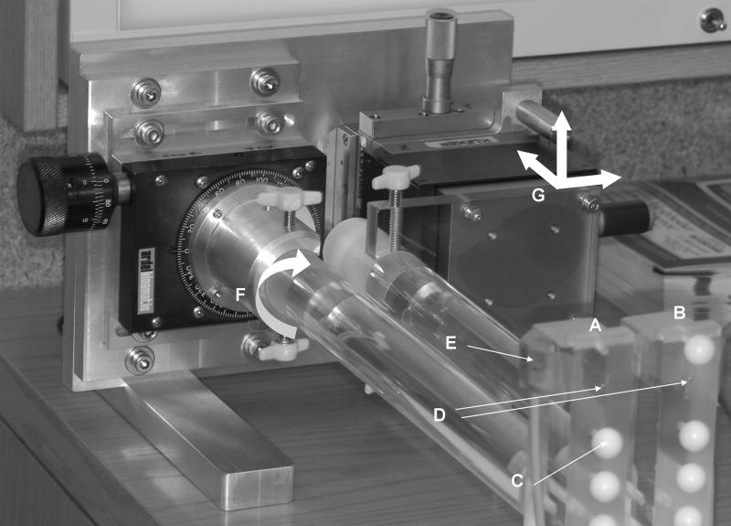
Figure 1.
Two plastic blocks (A, B) were constructed to represent the upper and lower jaw, and plastic spheres (C) 12 mm in diameter were attached to the blocks to represent the mandibular condyles. Three 0.8 mm diameter metal beads (D) were embedded in each block for identification and digitization of landmark points in three dimensions, during motion data collection and subsequent CT imaging. Plastic blocks were rigidly fixed to the calibration device and the sensors (E) were attached to the blocks via adhesive tape. The device provided for rotation about a single axis (F) and translation along three orthogonal axes (G).

