
Fig. 3.
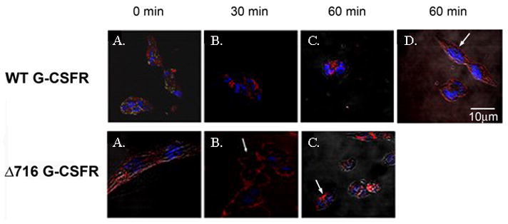
Internalization of the G-CSFR requires an intact ubiquitin-conjugation system. ts20 cells expressing the WT (upper panels) or Δ716 (lower panels) G-CSFR were stimulated with G-CSF (100 ng/mL) for 1h at 4oC then incubated at 30°C for 0 (A), 30 min (B), and 60 min (C). Cells were fixed, sequentially incubated with biotinylated antibody to the G-CSFR, streptavidin-Cy5, and Hoechst nuclear stain, then examined by confocal microscopy. (A-C) Cellular distribution of WT and Δ716 G-CSFR (red) and nuclei (blue). (D) WT G-CSFR-expressing cells incubated at 42°C (non-permissive temperature) for 60 min. Arrows indicate predominant membrane localization of the WT G-CSFR when ubiquitination is inhibited at 42– and of the Δ716 G-CSFR at the permissive temperature.