
Figure 1. Ribbon structure of apoE3-NT.
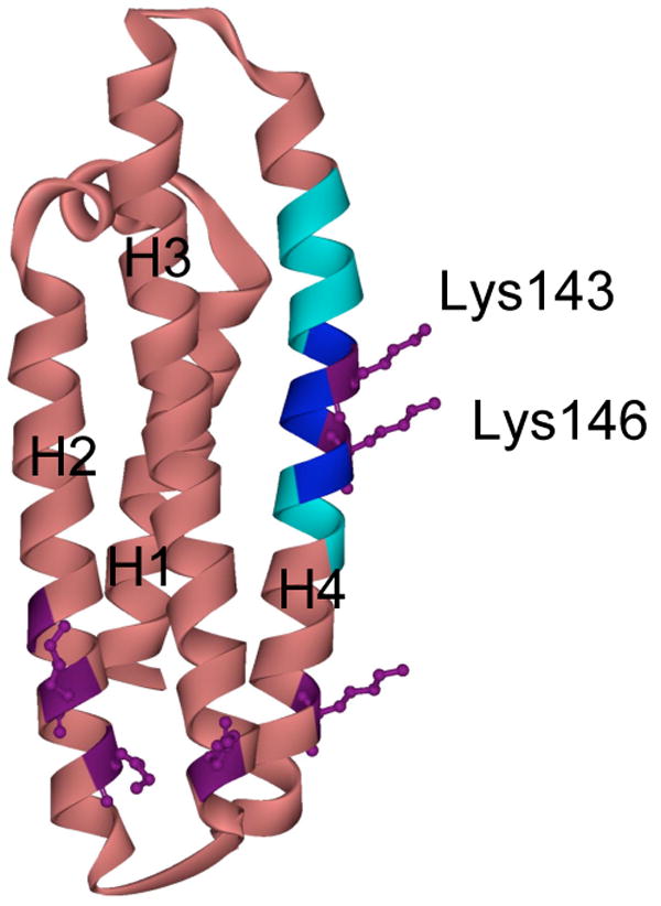
ApoE3-NT is comprised of 4 long amphipathic α-helices (labeled H1 to H4) that are folded into a helix bundle structure (rose) (18). The heparin binding site (dark blue) (residues 142-147, including Lys143 and Lys146 in purple) and the lipoprotein receptor binding site (light blue) (residues 136-150) are located on helix 4. The side chains of the other lysine residues visible in the crystal structure are indicated in purple.