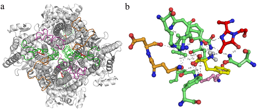
Figure 7.
Loops from three different monomers located around each active site of A. variabilis PAL double mutant. (a) The bottom view of A. variabilis PAL with loop 74–96 (monomer A, green), 436–458 (monomer A, green), 291–311 (monomer B, orange) and 394–419 (monomer C, magenta) are highlighted. (b) Networking among residues from three monomers in each active site with MIO (red) and docked Phe (yellow).

