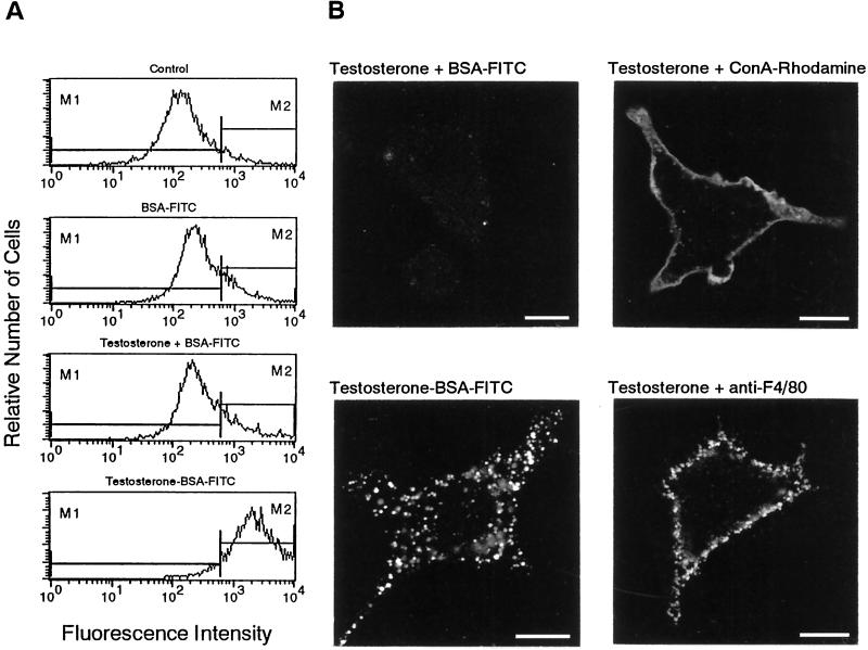
Figure 7.
Selective internalization of surface binding sites of testosterone. (A) Flow cytometry of IC-21 cells treated with the indicated substances for 15 min. (B) CLSM of cells incubated for 15 min with the indicated substances and the corresponding FITC-labeled secondary antibody. Note the difference between the smooth uniform surface labeling with Con A-rhodamine and the granular surface fluorescence of the F4/80 antigens. Bars, 10 μm.

