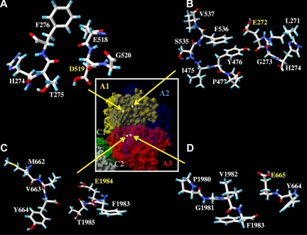
Figure 6.
Residues surrounding Asp519, Glu272, Glu1984, and Glu665. Factor VIII surface models of indicated regions based on the A domain homology model19 are drawn by Swiss PDB viewer; A1 domain (residues 1-336), A2 domain (residues 373-711), and A3 domain, (residues 1690-2332). Hydrogen, carbon, oxygen, sulfur, and nitrogen are colored cyan, white, red, yellow, and blue, respectively. There are no possible hydrogen acceptor or donor from the residues near the residues Asp519 (A), Glu272 (B), Glu1 984 (C), and Glu665 (D). (Inset) Factor VIII surface model of individual domains are drawn by Swiss PDB viewer and colored as yellow (A1), transparent blue (A2), red (A3), green (C1), and gray (C2). White dots indicate the location of side chain atoms of the indicated residues (Asp519, Glu272, Glu1984, and Glu665) as shown in the panels A, B, C, and D, respectively.

