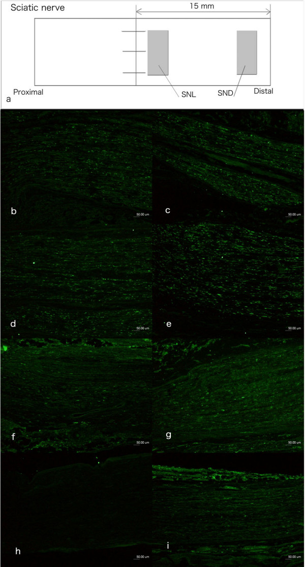
Figure 2.

Immunocytochemical staining of ATF3 in non-neuronal cells in the sciatic nerve. The non-neuronal cells were interpreted to be mainly Schwann cells based on the shape of the nucleus and location within basal lamina (also stained for S-100). The schematic drawing in (a) showed the sites where ATF3 stained cells were analysed in sciatic nerve of rats. SNL: distal site adjacent to the sutured lesion; SND: 15 mm distal to the sutured lesion. The number of cells was also calculated 15 mm proximal to and at the proximal site of the sutured lesion (results not shown). The photos on the left column (b, d, f) are from the site of lesion (SNL) and in the right column are from the distal nerve segment (SND; c, e, g, i) where the sciatic nerve was immediately repaired (b, c), repair after 30 days (d, e), 90 days (f, g) and 180 days (i). The contralateral uninjured sciatic nerve showed no or single ATF3 stained cells (h). Scale bar = 50 μm.
