Figure 6.
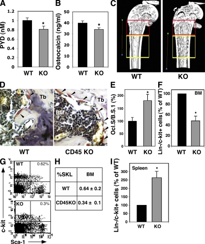
Reduced osteoclast function in CD45KO mice is associated with elongated trabecular zone and irregular localization of progenitors. (A) Plasma levels of PYD indicating bone resorption in WT and CD45KO. (B) Plasma levels of osteocalcin in WT and CD45KO mice. Values represent the mean ± SE (*, P < 0.05). (C) Three-dimensional constructions of the distal femurs of WT and CD45KO mice. The red frame indicates the metaphyseal region, near the bone growing plates, and the yellow frame indicates a region 1 mm ahead toward the diaphysis (see Materials and methods). Bars, 2 mm. (D) TRAP staining of femoral bone sections. Osteoclasts stained in red (arrowheads) are shown along the metaphyseal trabecules (Tb). (E) The ratio between osteoclast surface and bone surface (Ocl.S/B.S). Data are presented as the percentage of WT cells ± SE (*, P < 0.05). (F–H) Flow cytometry analyses of BM immature Lin−/c-Kit+ progenitors (F), presented as the percentage of WT (mean ± SE), and the more primitive SKL stem cells. A representative FACS plot (G) and summary (H) indicate lower levels of these primitive cells in the BM of CD45KO mice. *, P < 0.01. (I) Flow cytometry analyses of spleen-derived immature Lin−/c-Kit+ progenitors, presented as the percentage of WT (mean ± SE). Numbers indicate higher levels of these primitive cells in the spleen of CD45KO mice, *, P < 0.01.

