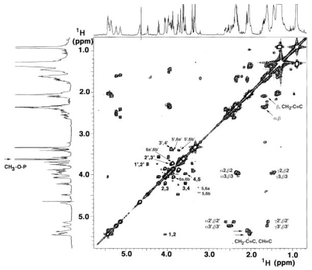
Fig. 4. 1H-1 H COSY of L. interrogans serovar Pomona lipid A at 600 MHz.
The 2-mg lipid A sample was dissolved at 25 °C in 0.6 ml of CDCl3, CD3OD, D2O (2:3:1, v/v/v). The numbering scheme is shown in Fig. 7A. The arrow at the left highlights the doublet near 3.61 ppm that arises from the methylated 1-phosphate. The two arrows at the lower right highlight the strong cross-peaks between the olefinic and vinylic protons of the secondary acyl chains.

