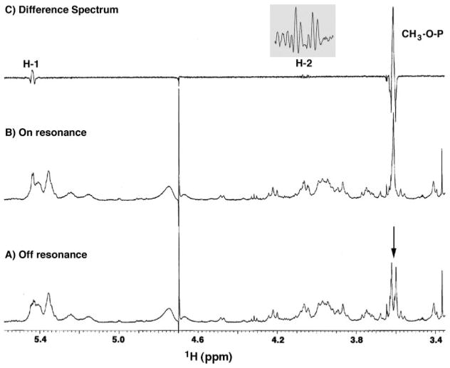
Fig. 5. Selectively 31P-decoupled 1H NMR spectra of L. interrogans serovar Pomona lipid A.
The sample used in Fig. 4 was subjected to 1H NMR analysis with the decoupler either off resonance or on the 31P atom resonance. The doublet at 3.61 ppm (arrow in A) collapses to a singlet (B) when the phosphorus atom is selectively irradiated. The difference spectrum (C) highlights additional coupling of the phosphorus atom to the H-1 and H-2 signals of the proximal sugar. The inset in C shows the H-2 difference spectrum in detail.

