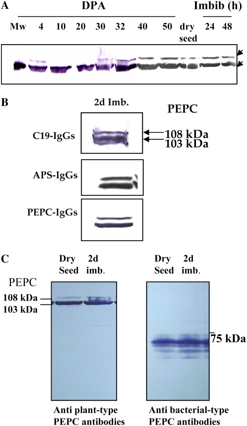
Figure 2.
Immunocharacterization of PEPC. A, At the indicated times, soluble proteins (200 μg) of nondesalted extracts from whole seed were separated by SDS-PAGE (10% acrylamide), transferred onto nitrocellulose, and probed with polyclonal antibodies raised against sorghum leaf PEPC. The arrows indicate 108- and 103-kD PEPC subunits. B, The 103- and 108-kD polypeptides are intact and classical plant-type PEPC (PTPC) subunits. The experiment was performed as in A and probed with specific C19-IgGs, APS-IgGs, or anti-sorghum leaf PEPC IgGs (30 μg per 20 mL of incubation medium). C, The experiment was performed as in A. The transferred protein from dry and 2-d-imbibed seeds was incubated with polyclonal anti-sorghum leaf PEPC IgGs (30 μg per 20 mL of incubation medium) or antibacterial-type PEPC4 IgGs from Arabidopsis (20 μg per 20 mL of incubation medium). Immunolabeled proteins were detected by a horseradish peroxidase assay. [See online article for color version of this figure.]

