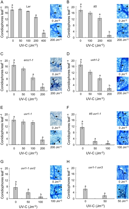
Figure 4.
Effects of UV shielding and DNA repair defects on UV-induced resistance to H. parasitica. Leaves of Ler wild-type (A), shielding defective (B), NER defective (C–E), shielding and NER defective (F), NER and CDP photolyase defective (G), or NER and 6-4PP photolyase defective (H) plants were inoculated with HpHind4 24 h after mock treatment or UV exposure, and the number of conidiophores per leaf was determined 7 d after inoculation. On the right of each section, representative samples of leaves that were inoculated with HpHind4 24 h after mock UV treatment or UV irradiation, incubated for 7 d, and then stained with lactophenol-trypan blue are shown. Hyphae-bearing haustoria or HR lesions are indicated by black arrows or arrowheads, respectively. Bar in A (bottom right) = 50 μm. [See online article for color version of this figure.]

