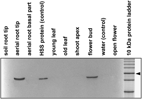
Figure 1.
Expression analysis of HSS in various tissues of Phalaenopsis. Soluble protein (20 μg) extracted from the tissues was separated by SDS-PAGE and blotted onto a polyvinylidene difluoride membrane. As a positive control, 10 ng of purified HSS protein was applied. Detection was performed with affinity-purified antibody against HSS of Phalaenopsis. Soil roots were roots that penetrated the substrate, whereas aerial roots did not. Young and old leaves were approximately 3 and 10 cm long, respectively. The 50-kD band of the marker protein is labeled with an arrowhead.

