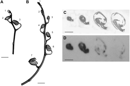
Figure 5.
Inflorescences of Phalaenopsis plants. A and B, The numbers labeling the buds refer to the tracer feeding experiments shown in Table I. C and D, Tissue prints of longitudinal sections of Phalaenopsis buds on nitrocellulose membrane developed with affinity-purified antibody against HSS of Phalaenopsis (D) and stained for total protein by Indian ink (C). Size bars in A to D = 1 cm.

