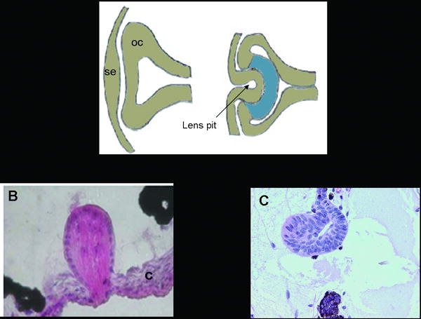
Figure 2.

Comparison of lens development with two methods of lens regeneration. A. Schematic showing lens development, which involves a series of inductive interactions between the surface ectoderm and the optic cup. The lens pit eventually gives rise to the lens. se: surface ectoderm, oc: optic cup. B. Lens regeneration in Xenopus laevis. The regenerated lens comes from transdifferentiation of cells in the outer cornea (c). C. Lens regeneration in the newt. The regenerated lens comes from transdifferentiation of cells in the dorsal iris.
