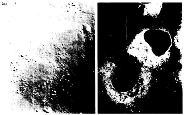
FIG. 2.
Confocal microscopy of permeabilized UOK-123 cells. Cytospin preparations of UOK 123 cells were permeabilized and stained with monoclonal antibody HC 10, which binds to unfolded class I heavy chains. Immunofluorescence images (right panel) demonstrated a reticular staining pattern in the cytoplasm consistent with reactivity in the endoplasmic reticulum. No surface staining was observed. A phase-contrast image of the same field is shown in the left panel.

