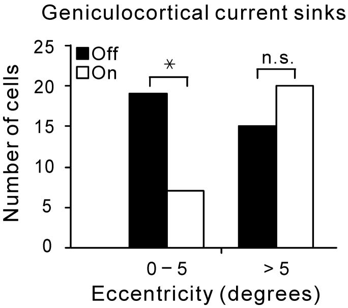
Figure 3. Off-center geniculate cells dominate the representation of the area centralis in cat visual cortex.

a Near the cortical representation of the area centralis (< 5 degrees eccentricity), current sinks generated by off-center geniculate afferents were more frequently found than current sinks generated by on-center geniculate afferents (P = 0.02, Chi-square test, data obtained from 9 cats). This difference was not found outside of the area centralis. b. Recordings from LGN demonstrate that on- and off-center geniculate cells are balanced in number at two different eccentricity ranges. Significance was assessed with a Chi-square test (n.s.: not significant; *: P = 0.018).

