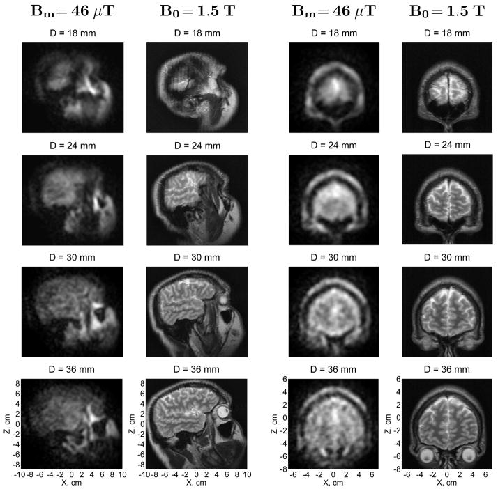
Fig. 3.
Microtesla MRI of the human head compared to conventional MRI. The 3D ULF MR images of the head side and the forehead area were acquired at 46 μT measurement field. Each image in the figure represents a 6 mm-thick layer of the head. D is the depth of the central plane of a given layer with respect to the bottom of the cryostat. The in-plane resolution is 3 mm × 3 mm. The high-field 3D image of the same subject’s head was acquired by conventional MRI at 1.5 T. Each high-field image in the figure corresponds to the same layer of the head as the ULF image on its left.

