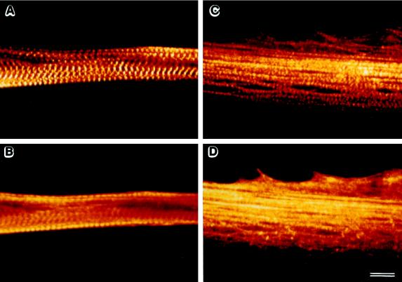
Figure 7.
Rac1V12 inhibits the organization of α-actinin and myosin into sarcomeres. QMb-LA29 stably expressing neomycin resistance (A and B) or Rac1V12 (C and D) were cultured in DM at 41°C for 3 d and then processed for immunofluorescence. Confocal immunofluorescence micrographs of myotubes double labeled for α-actinin (A and C) and myosin (B and D) show that myotubes expressing Rac1V12 are flattened onto the substrate and that both α-actinin and myosin are organized in bundle-like assemblies, lacking both lateral alignment and periodic cross-striation, which are easily detected in controls. The two chromophores are depicted with the same color scale in panels A and B and panels C and D. Bar, 10 μm.

