
Figure 1.
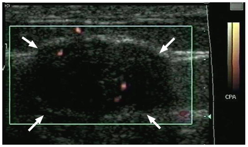
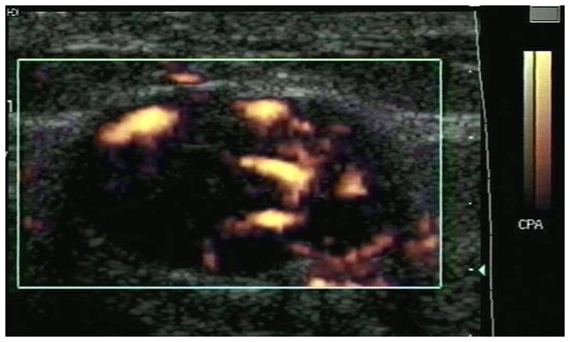
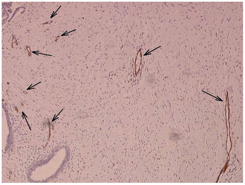
Example of a fibroadenoma (arrows) imaged in power Doppler mode pre (A) and post (B) injection of 10 ml of Levovist. A pathology specimen obtained from the same lesion is presented in (C) with areas stained with CD31 shown in brown (arrows).