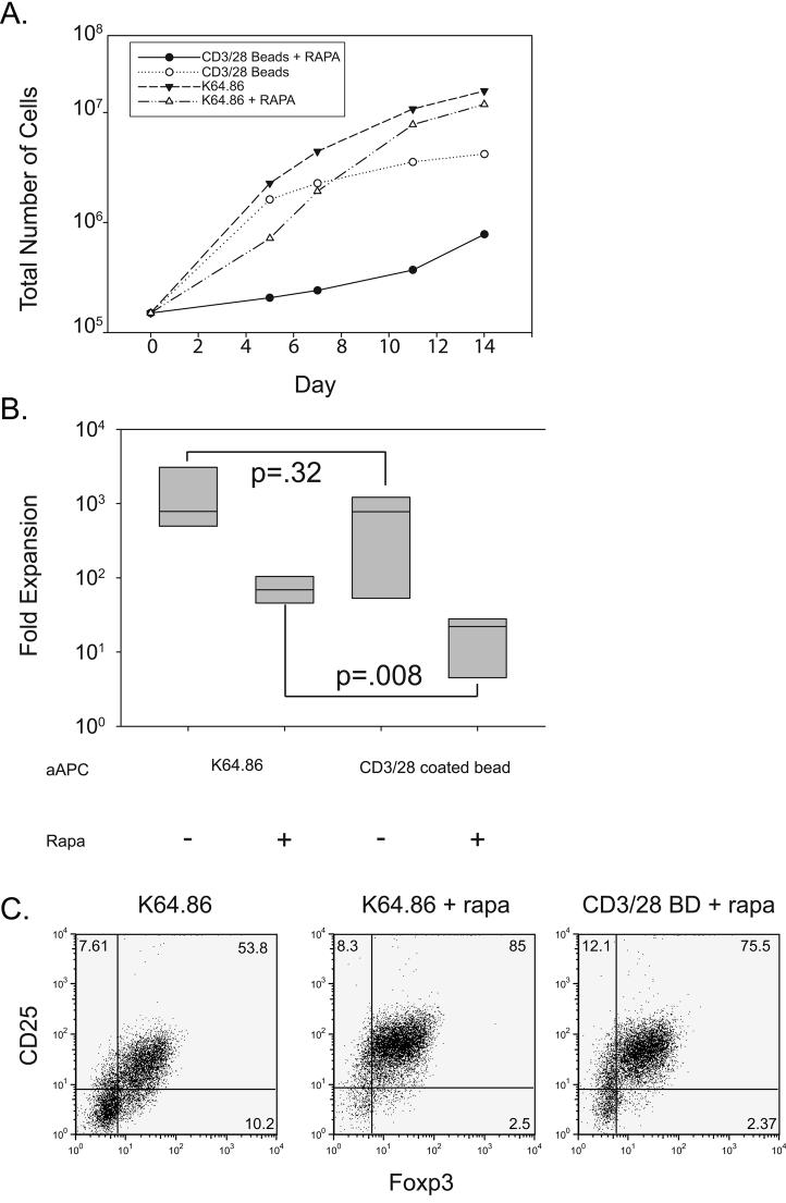
Figure 2. CD28 ligands on cell-based aAPCs expand Tregs more efficiently than bead-based aAPCs.
A. 2×10⁵ enriched Tregs were stimulated with either CD3/28 Ab coated beads or anti-CD3 loaded K64.86 aAPCs and cultured in the presence or absence of rapamycin for two weeks. Expansion was measured as described in the Materials and Methods. B. Box plot showing fold expansion for each culture condition using data collected from four donors. Fold expansion was determined by dividing the total number of cells at the end of culture by the initial starting number stimulated by the indicated aAPC in the presence or absence of rapamycin. C. Analysis of Foxp3 and CD25 expression in Tregs expanded with either CD3/28 Ab coated beads (BD) or anti-CD3 loaded K64.86 aAPCs. Data is representative of four independent experiments.

