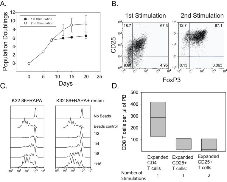
Figure 7. Restimulation of T regulatory cells enables greater than 1000 fold expansion without loss of in vitro or in vivo suppressive activity.

A. 2×10⁵ enriched Tregs were stimulated by K64.86 aAPCs and expanded in the presence of rapamycin. Population doubling rate was measured by cell counting. Each data point represents the average of three independent experiments (error bars represent standard deviation). When cell expansion reached the plateau phase, the culture was restimulated with K64.86 cell-based aAPCs (open circles) or left alone (filled in circles). B. Analysis of Foxp3 and CD25 expression in enriched Tregs stimulated with K64.86 aAPCs once or twice in the presence of rapamycin after 14 days of culture. Data is representative of four independent experiments. C. The in vitro suppressive activity of cell populations shown in A and B was measured by mixing CFSE-labeled autologous PBMCs, anti-CD3 Ab coated beads, and the indicated ratio of expanded Tregs and PBMCs. After 4 days of culture, CFSE dilution was measured by flow cytometry. D. 2×10⁶ cells from each culture shown in A. were mixed with 1×10⁷ autologous PBMCs and injected into NOG mice (6 mice per group). After 8 weeks, the mice were bled and the number of human CD8 T cells/μl of blood was determined.
